


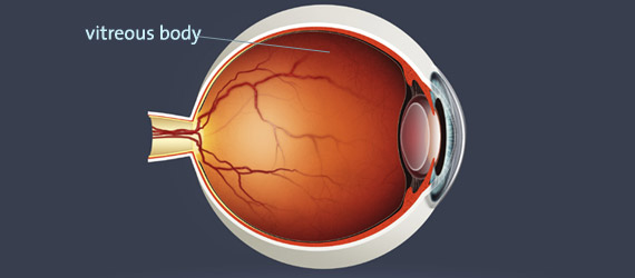
What is the vitreous body and what are vitreous diseases?
These diseases affect the posterior part of the eye, the vitreous body (corpus vitreum). This gel-like, transparent substance has the function of “stabilizing” the posterior part of the eye.
Learn more about vitreous body diseases in this 3-minute video:
How do you notice these diseases?
Your symptoms could be the following:
- You notice haze, streaks, flashes of light or floaters (vitreous symptoms).
- You are sensitive to light and suffer from glare problems, see halos or a shadow.
- You see a shadow, notice a deterioration of your vision or you have similar symptoms (indication for possible vitreous opacities or vitreous hemorrhage, retinal tears or retinal detachment).
If you have these or similar symptoms, a prompt and careful eye examination is required to clarify the exact cause.
Examination of the vitreous body
To carefully examine the posterior part of your eye, a (painless) dilation of the pupil is necessary. Therefore, please leave your car at home. Use public transportation or take a “driver” along. The pupil dilating drops will affect your ability to drive for approx. 4 hours.
Depending on the results of the eye examination performed, a surgery may or may not be necessary.
In a comprehensive conversation, I will advise you about the necessity of surgery, the details of the procedure, the expected surgical outcome and possible side effects. It is a major concern for me to address all of your questions and to explain in detail how the surgery will be performed.
Even vision impairing or subjectively disturbing vitreous opacities (such as for example a synchysis scintillans) can be treated with surgery.
The vitrectomy (vitreous surgery)
Vitrectomy works in a similar way as endoscopy. This means making tiny holes into the interior of the eye and removing the vitreous body. As a replacement for the vitreous body, the eye is filled with liquid. Depending on the diagnosis, the retina may be treated at the same time.
In case of more severe diseases an additional scleral buckling procedure may be performed. A “sponge” material is stitched onto the eye ball to create a “buckle” on the eye wall.
If you have signs of a cataract formation, I will perform the vitrectomy surgery in combination with a cataract procedure. If only vitrectomy is performed, your lens will develop cataract formation within the next 5 years leading to deterioration of your usual visual acuity. Thus, another surgery would become necessary. However, a combined surgical procedure prevents a second operation within the next 1 to 5 years. International investigations as well as my own prospective studies have shown safe and excellent results after combined surgery.
Vitrectomy (as well as scleral buckling surgery) is performed under general anesthesia. Please expect to stay 1 to 2 nights in the clinic. Avoid swimming, sauna and similar activities after the surgery for approx. 2 to 3 weeks, physical stress and sports for approximately 3 to 4 weeks.
I perform vitrectomy with the newest sutureless microincision techniques. The cannulas are extremely thin, they range from 25 gauge (≈ 0.5 mm) to 27 gauge (≈ 0.4 mm). The results are excellent and I have thoroughly studied these techniques in many research projects: worldwide, it is the safest method available.
These incisions are so small by now that they are self-healing and usually don’t require sutures for wound closure.
This means for you: no pain, less scratching and no foreign body sensation in your eye, less redness, as well as a shorter hospital inpatient stay and faster recovery.
During vitrectomy, I also occasionally use a YAG laser. YAG stands for “yttrium aluminum garnet”, which is the material the laser crystal is made of. The use of the laser makes it possible to “weld” retinal holes or tears with pinpoint accuracy. The laser creates tiny scar tissue around the lesion and this helps to fix the retina to its substrate and prevent further detachment.
I have successfully performed hundreds of vitrectomies, so you can benefit from my years of experience.
Should you have questions about vitreous diseases or surgical procedures, please contact me.